What Is a Retinal Detachment?
The retina is a thin, light-sensitive layer lining the inside back of the eye. For it to work, it must remain in contact with the underlying tissue that nourishes it. A retinal detachment occurs when the retina lifts or separates from this supporting layer. When that happens, the affected area stops working — often described as a curtain, shadow or dark area moving across the vision.
A common pathway begins with the vitreous, the gel that fills the eye. With age (and earlier in near-sighted eyes or after injury), the vitreous can shrink and pull away from the retina — a posterior vitreous detachment (PVD). Usually harmless, a PVD can sometimes tug hard enough to create a retinal tear. Fluid can then pass through the tear and accumulate behind the retina, separating it. This is called a rhegmatogenous detachment, the most common type. Other mechanisms — traction from scar tissue, or fluid accumulating beneath an intact retina — occur in specific conditions such as advanced diabetic eye disease or inflammation.
Retinal detachment is an emergency in the truest sense: the longer the retina remains detached, the more its cells are deprived of support, and the less vision may recover after successful repair.
Warning Signs That Must Not Wait
Learn these signs, and treat them as a same-day problem rather than a "wait and see" symptom:
- A sudden shower of new floaters — many dark spots or a "cobweb" appearing at once
- Repeated flashes of light, especially in peripheral vision or in the dark
- A curtain, shadow or veil descending over part of the visual field
- Sudden, painless reduction or loss of vision
None of these proves a detachment — but all of them justify urgent dilated retinal examination. If a detachment is found, the discussion shifts immediately to timing and repair options. Do not wait for an online reply, and do not drive yourself if your vision is significantly affected.
How the Diagnosis Is Made
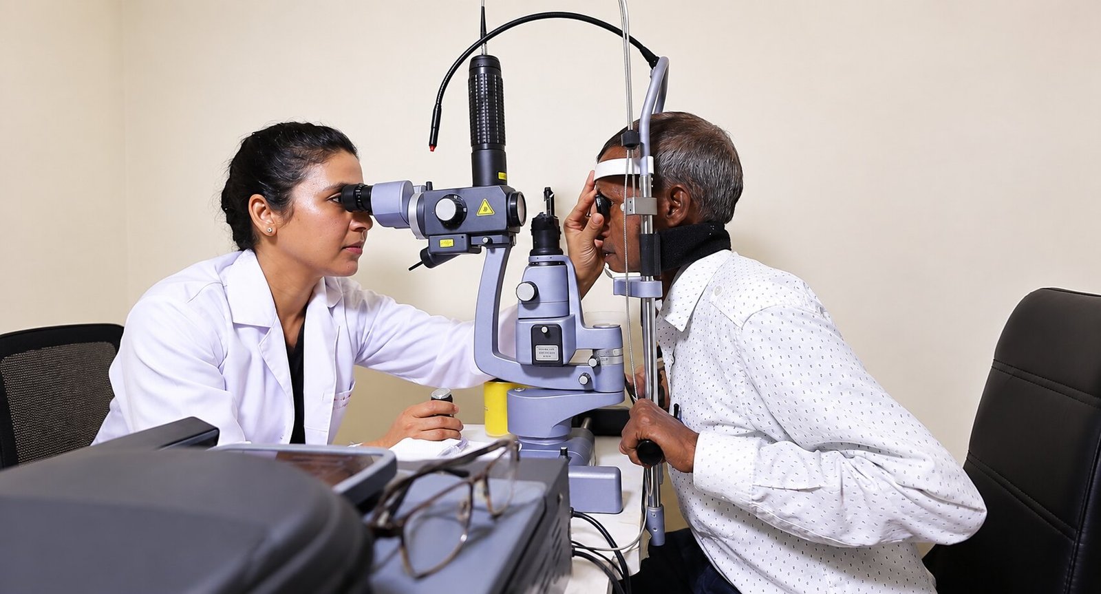
After a focused history, the retina is examined with a dilated fundus examination. The specialist looks for tears, the extent and height of any detachment, whether the macula is involved (a key factor in visual prognosis), and any complicating features such as vitreous haemorrhage.
Imaging supports the picture: B-scan ultrasound is invaluable when blood or media opacity blocks the view, and OCT helps assess the macula. Findings are explained immediately — with detachment, clarity and speed both matter — and the planned approach, alternatives and risks are discussed before consent.
How Retinal Detachment Is Treated
Treatment depends on the type, size, location and duration of the detachment, and on the specific eye. Broadly:
- Retinal tears without detachment are often sealed with laser retinopexy or cryotherapy in an outpatient setting, creating a scar barrier that prevents fluid passing behind the retina.
- Scleral buckling — a silicone element is placed around the eye wall, gently indenting it against the retinal break so the retina reattaches as fluid is drained or resorbed.
- Vitrectomy — the vitreous gel and any traction are removed, the retina is repositioned, and a gas bubble (or occasionally silicone oil) is placed to support healing while the laser seals the breaks. Specific head-positioning and air-travel restrictions, where needed, are given as individual surgeon instructions — they are never universal rules.
- Pneumatic retinopexy — selected shallow detachments with a single superior break may be treated with an in-office gas bubble plus laser.
Each option carries benefits, limitations and risks — including cataract formation, raised eye pressure, infection, or redetachment — and these are discussed openly. The goal is reattachment with the best achievable functional recovery; individual outcomes vary with macular involvement and duration.
Recovery and Follow-Up
Recovery involves positioning instructions where a gas bubble is used (these vary — follow your personal written plan), activity limits, and strict attendance at follow-up visits where the retina is re-examined and the bubble's absorption is monitored. A new shower of floaters, flashes, an enlarging shadow, increasing pain or redness warrants immediate review.
Because the fellow eye shares risk factors, it is examined carefully too — and any asymptomatic tears there can be treated before they become a problem. Long-term, people who have had a detachment benefit from periodic reviews and from knowing the warning signs above.
Common questions
Is retinal detachment painful?
Typically no — the classic symptoms (floaters, flashes, curtain, vision loss) are painless, which is why they are sometimes ignored. Pain is more suggestive of other problems such as inflammation or glaucoma, which also need urgent review.
Can a detached retina heal on its own?
No. Once separated, the retina will not reattach without treatment, and delay reduces the chance of good visual recovery — especially if the macula is involved.
How quickly must detachment be treated?
As soon as safely possible — typically within hours to days depending on macular involvement. Time matters because retinal cells deprived of support lose function the longer they remain detached.
Will my vision return to normal after surgery?
Outcomes vary. Detachments caught before the macula detaches often recover very well; long-standing macula-off detachments may retain some central deficit. Your surgeon will give an honest, individual outlook.
What causes the flashes and floaters before a detachment?
They usually reflect the vitreous gel separating from the retina and tugging on it. A thorough dilated examination distinguishes a harmless PVD from a tear — the two can feel identical.
Am I at higher risk if I am highly near-sighted?
Yes — longer eyes are more prone to early vitreous separation and retinal tears. Regular dilated examinations are especially worthwhile.
Can I fly after retinal detachment surgery?
Only when your surgeon confirms it is safe — generally after any intraocular gas has fully absorbed, since cabin pressure changes affect the gas bubble. Never fly against your surgeon's advice.
What is the success rate of repair?
Most detachments are successfully reattached, but 'anatomical success' and 'visual recovery' are different measures, and redetachment can occur. Your individual risk profile is discussed before surgery.
Is scleral buckle or vitrectomy 'better'?
Neither is universally better; each suits different detachments. Factors include break location, lens status, proliferative changes and surgeon judgement — this is exactly what consultation is for.
What should I do right now if I see a curtain over my vision?
Arrange an urgent dilated retinal examination today — contact the hospital or a nearby eye emergency service. Do not wait for an online response, and avoid driving if your vision is affected.