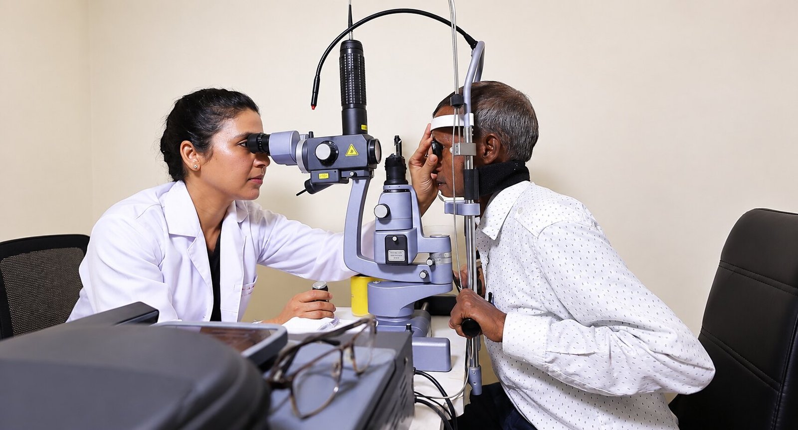
About Intermediate and Posterior Uveitis
['\nIntermediate uveitis involves the vitreous (often appearing as persistent floaters or haze), while posterior uveitis involves the choroid and retina — sometimes painless, discovered only when vision dims. Infectious causes (including toxoplasmosis and TB, region-dependent) must be distinguished from autoimmune patterns, because steroids — so useful in the latter — can worsen the former.
\nEvaluation combines examination, OCT, angiography and targeted systemic testing, with treatment ranging from local and systemic steroids to steroid-sparing immunosuppression in chronic disease, plus specific anti-infective therapy where indicated. Multidisciplinary coordination is routine. Follow-up is long-term by design: quiet eyes still harbour activity that only imaging reveals.
\n']Common questions
Why did I get floaters suddenly?
Vitreous inflammatory cells are a classic intermediate-uveitis sign; new floaters always justify examination.
Is posterior uveitis always visible as a red eye?
No — the back of the eye can be heavily inflamed with a white, comfortable eye. Vision change is the clue.
Will I need immunosuppression?
Chronic or vision-threatening non-infectious disease often does best with steroid-sparing agents, chosen and monitored jointly with physicians.
Can infections cause this?
Yes — which is exactly why treatment never precedes a structured infectious work-up in typical practice.
How long is follow-up?
Often years, tapering in frequency as durable control is proven.