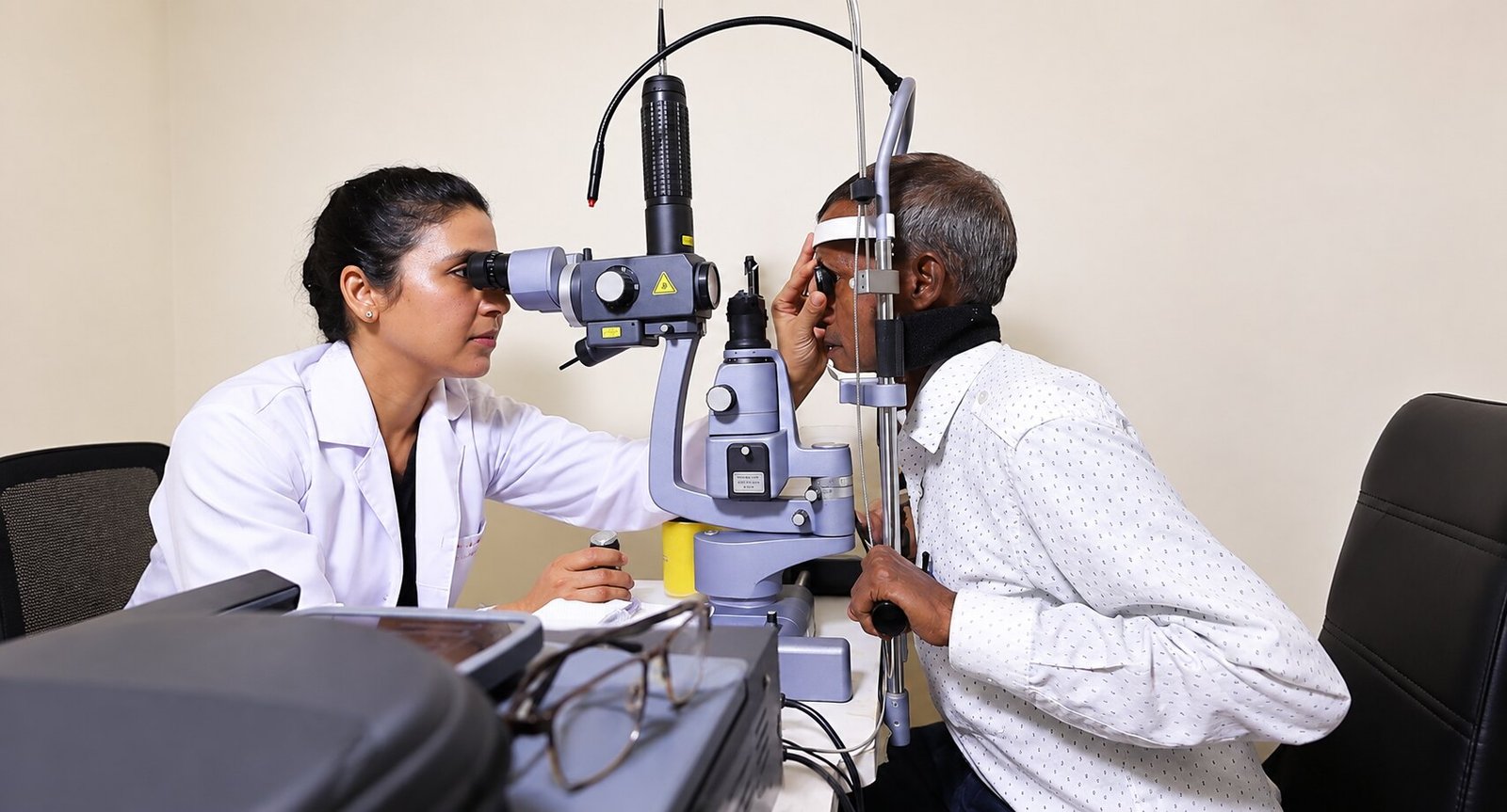
Understanding Diabetic Retinopathy
Diabetic retinopathy is a complication of diabetes that affects the retina — the light-sensitive layer at the back of the eye. Over time, elevated blood sugar can weaken the walls of the tiny retinal blood vessels, causing them to leak fluid, bleed, or close off. The retina depends on a precise, healthy blood supply, so these changes can gradually threaten vision.
The condition is broadly grouped into stages. In non-proliferative diabetic retinopathy (NPDR), small vessel changes — microaneurysms, tiny haemorrhages, leakage or vessel closure — may be seen. Vision can remain normal for years, which is exactly why screening matters: damage can progress silently. In proliferative diabetic retinopathy (PDR), the retina responds to poor blood supply by growing fragile new vessels, which can bleed into the vitreous (vitreous haemorrhage) and pull on the retina, raising the risk of retinal detachment.
When leakage affects the macula — the central part of the retina responsible for detailed vision — it is called diabetic macular oedema (DME). DME is one of the most common reasons people with diabetes notice blurred or distorted central vision.
Why Timely Screening Matters So Much
Diabetic retinopathy often causes no symptoms in its early stages. Many people only learn they have it during a routine dilated eye examination. By the time vision blurs, changes may already be significant. Evidence consistently shows that early detection, together with good systemic control, offers the best chance of preserving vision.
How often screening is recommended depends on your type of diabetes, duration, pregnancy status and other factors — your eye specialist and physician will advise a schedule that fits you. The key message is simple: do not wait for symptoms. If you have diabetes, a dilated retinal examination is part of your overall diabetes care, in the same way that kidney and foot checks are.
Eye treatment never replaces diabetes management. Controlling blood sugar, blood pressure, cholesterol and kidney health — together with your physician — remains the foundation of protecting your sight.
How Dr. Neha Mohan Evaluates Diabetic Eyes
Every evaluation begins with history: the type and duration of diabetes, recent HbA1c and blood-pressure readings, kidney status, medications (including any blood thinners), previous laser or injections, and how your vision feels day to day. This context shapes both the examination and the plan.
A dilated retinal examination allows a direct view of the peripheral and central retina. Depending on findings, imaging may be recommended:
- OCT (optical coherence tomography) — shows cross-sectional detail of the macula, revealing swelling, fluid pockets and response to treatment.
- Fundus photography — documents the retina so changes can be compared visit to visit.
- Fundus fluorescein angiography (FFA) — maps areas of leakage and poor blood supply, often guiding laser planning.
- B-scan ultrasound — used when bleeding inside the eye prevents a direct view of the retina.
Findings are explained in plain language, and you will see and discuss your own images where helpful. Staging is described honestly — many eyes need monitoring rather than immediate treatment, and that is a valid, planned decision, not "doing nothing."
Treatment and Monitoring Options
Management depends on the stage, the macula, and your overall health. Broad categories include:
- Observation with structured follow-up — for mild changes, with clear return precautions.
- Medical/systemic optimisation — closer diabetes, blood-pressure and lipid control with your physician, which genuinely slows progression.
- Retinal laser — focal or panretinal patterns may be used to treat leakage or to reduce the drive for abnormal new vessel growth.
- Intravitreal injections — anti-VEGF medicines, and sometimes steroids, are the mainstay for diabetic macular oedema in many patients. Treatment typically follows an initial loading phase and then a personalised interval — your plan is set by examination and OCT response, not by a fixed public schedule.
- Vitreoretinal surgery — considered for complications such as non-clearing vitreous haemorrhage or tractional retinal detachment.
Realistic goals are discussed openly: treatment aims to stabilise and often improve vision, but results vary between individuals, and some eyes respond more slowly than others. Attending follow-up even when vision feels stable is essential, because active disease can recur quietly.
Preparing for Your Visit and Living Well With Diabetes
Helpful preparation includes bringing your current spectacles, previous eye reports and scans, a list of medicines and allergies, recent diabetes and blood-pressure readings, and a companion if dilation may blur your vision for a few hours. These are suggestions, not requirements — come even if you cannot organise everything.
Between visits, seek urgent assessment for sudden vision drop, a shower of new floaters, flashes or a shadow. Otherwise, protect your sight the same way you protect the rest of your body: regular reviews, honest conversations with your care team, and never skipping prescribed medication without advice.
Common questions
Can diabetic retinopathy be cured?
There is no simple cure, but progression can often be slowed substantially and vision loss prevented or reduced with timely treatment plus good diabetes, blood-pressure and cholesterol control. Early-stage disease may only need structured monitoring.
My vision is fine — do I still need a retinal check?
Yes. Diabetic retinopathy frequently causes no early symptoms. Screening before symptoms appear is when treatment options are usually simplest and most effective.
What is the difference between diabetic retinopathy and macular oedema?
Retinopathy describes vessel disease across the retina; macular oedema is swelling of the central retina caused by leakage. An eye can have both, and each is assessed and treated differently.
Are injections painful?
Anaesthetic drops and careful preparation make the procedure brief and generally well tolerated. Mild redness or floaters can occur; serious complications are uncommon but explained before consent.
How often will I need treatment or follow-up?
It depends on the stage and response. Some patients need a course of injections with tapering intervals; others need annual checks only. Your schedule is individualised from examination and OCT findings.
Does controlling sugar reverse eye damage?
Good systemic control slows progression and reduces risk, but established retinal changes may still need laser, injections or surgery. Eye care and diabetes care work together — neither replaces the other.
Can diabetic retinopathy cause blindness?
If advanced disease is left untreated, severe vision loss is possible. Regular screening and timely treatment make this far less likely — which is why screening is emphasised so strongly.
Is laser treatment safe?
Retinal laser is a well-established procedure with defined benefits and risks, which are explained before consent. Side effects such as temporary blur or mild discomfort are discussed honestly.
Can I drive after a dilated examination?
Dilation commonly blurs near vision and increases light sensitivity for several hours, so bringing a companion or avoiding driving yourself is sensible.
Do blood thinners need to be stopped before injections or laser?
Never stop prescribed medication on your own. The team reviews your medicines and coordinates with your physician when any adjustment is clinically appropriate.