Retina Care in Delhi.
Clear Guidance at Every Step.
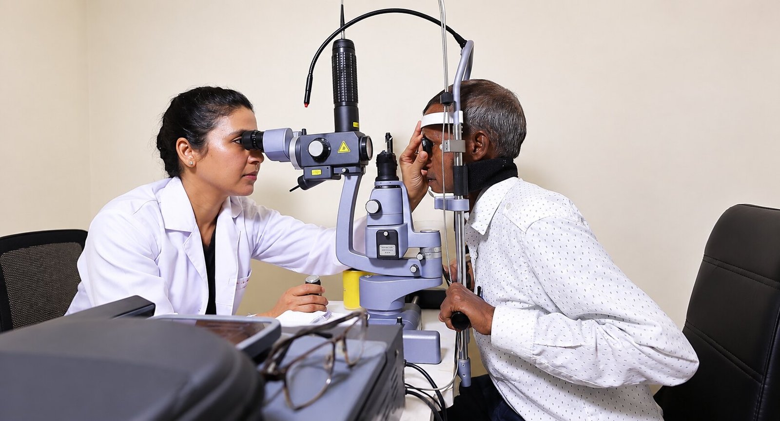
Detailed retinal evaluation, clear explanations of findings, evidence-based treatment options and planned follow-up — with coordination between patients, families, physicians and neonatal teams whenever required.
MBBS • MS Ophthalmology • FICO (UK) • FAICO • FLVPEI
Associated with Jain Eye Hospital & Laser Centre

How detailed retinal imaging helps map, interpret and monitor the back of the eye.
Feel prepared before you arrive.
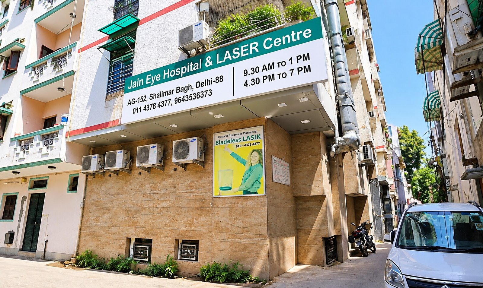
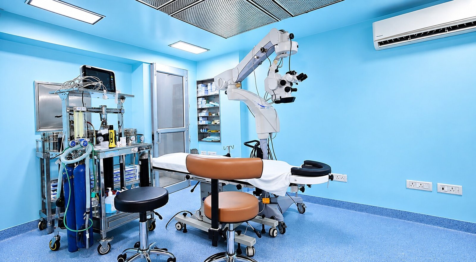
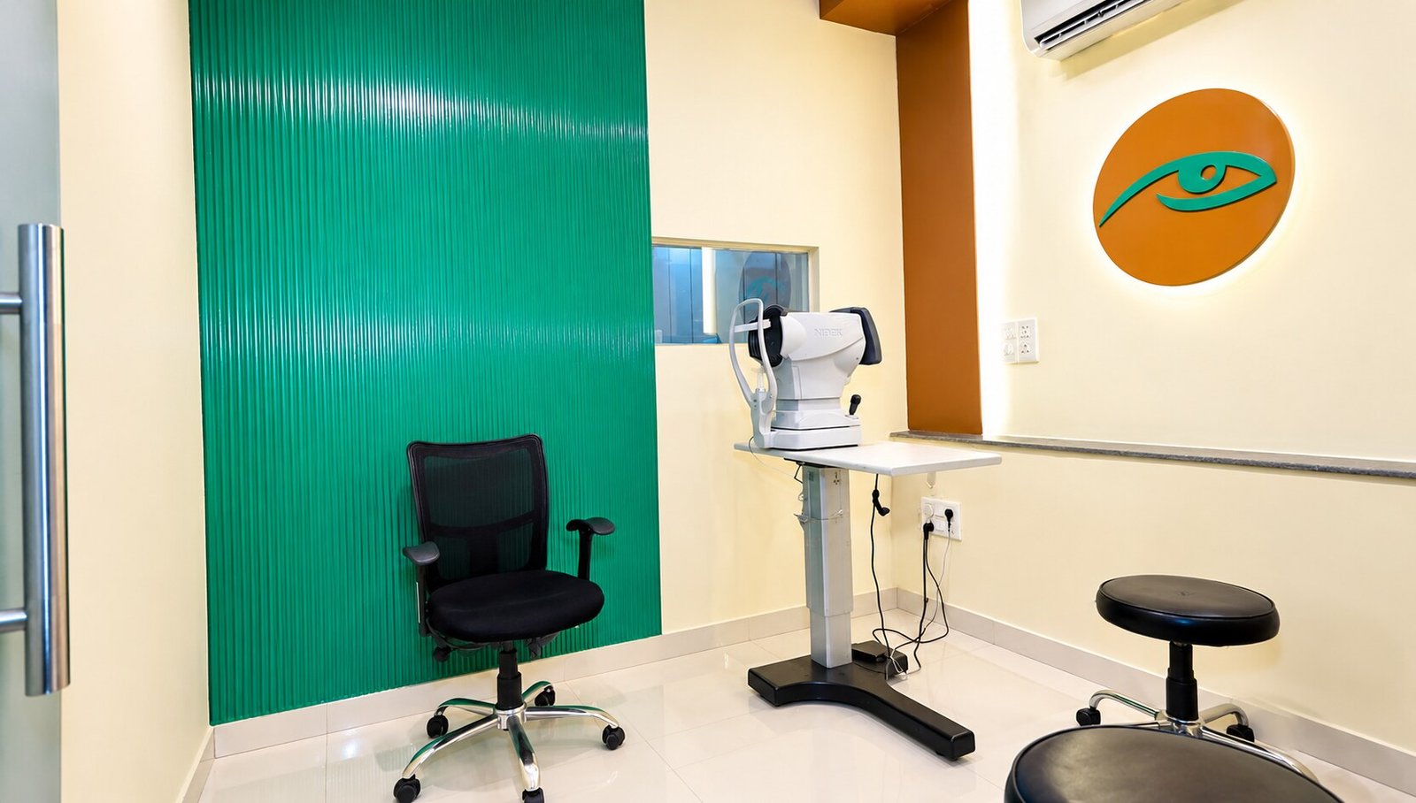
From the entrance and front desk to the examination and procedure rooms, get a clearer sense of the setting where your retina care journey begins.

Care Built on Detail, Explanation and Continuity
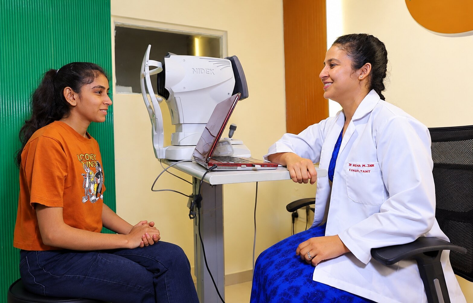
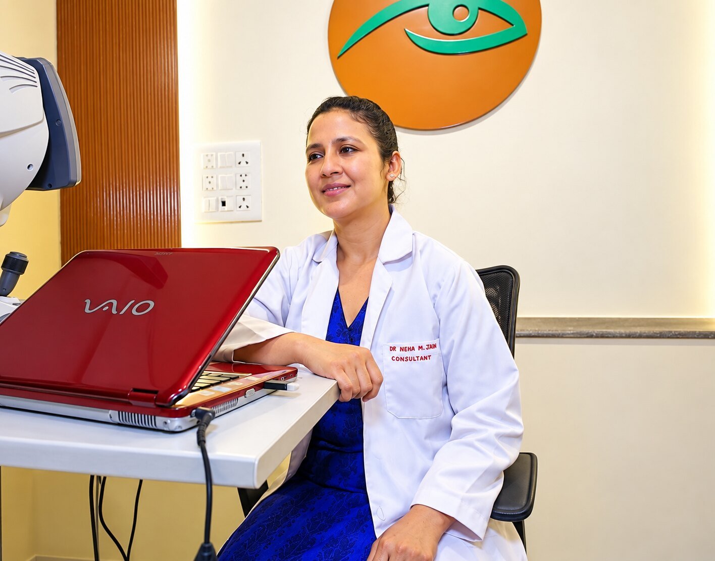
Dr. Neha Mohan is an ophthalmologist with focused training in vitreoretina, uvea, and retinal care. Her consultation approach centres on detailed examination, clear explanation of retinal findings, evidence-based options, careful follow-up, and coordination with patients, families, physicians, paediatricians, and neonatal teams whenever required.
- Careful, unhurried assessment of retinal and uveal conditions
- Findings explained in plain language with imaging where helpful
- Observation, medical, laser, injection and surgical options discussed as appropriate
- Coordinated care with physicians, paediatricians and neonatal teams when needed
- Planned follow-up with written recovery guidance
Three Focused Areas of Expertise
A short visual guide to how retinal imaging supports examination, mapping, explanation and follow-up. It is educational, never a diagnosis.
Retina & Vitreous
Diabetic retinopathy, retinal detachment, macular disorders, retinal laser and vitreoretinal surgery — evaluated and managed with careful staging and honest counselling.
Explore Retina Care →
Uvea / Uveitis
Inflammation inside the eye needs structured evaluation to look for underlying causes — with clear warnings against unsupervised steroid use and close multidisciplinary coordination.
Explore Uveitis Care →
Retinopathy of Prematurity
Compassionate, time-sensitive retinal screening for premature babies, coordinated with paediatric and neonatal teams — explained calmly for parents at every step.
Explore ROP Care →Retina Symptoms That Should Never Wait
This section is educational, not a diagnostic tool. Some retinal conditions begin with few or no symptoms — but the signs below can indicate problems that benefit from timely assessment.
Sudden Flashes of Light
New or repeated flashes, especially in low light, can accompany changes in the vitreous gel or retinal traction.
A Shower of New Floaters
A sudden increase in floaters, dots or cobweb-like shapes may suggest a retinal tear and warrants prompt examination.
A Curtain or Shadow
A dark curtain, shadow or veil moving across part of the vision can be a sign of retinal detachment — seek urgent care.
New Distortion
Straight lines appearing wavy or a central dark patch can relate to macular conditions and should be assessed.
Sudden Vision Loss
Unexplained, rapid reduction in vision — in one or both eyes — needs same-day professional evaluation.
Symptoms After Eye Injury
Floaters, flashes, pain or reduced vision after trauma require examination even if the injury seemed minor.
Seek Urgent Eye Assessment
For sudden vision loss, a curtain/shadow, or a shower of floaters, seek urgent eye assessment immediately — do not wait for a website reply.
Retinal Changes Can Begin Before Any Symptom
Diabetes can affect the fine retinal blood vessels long before vision changes. Regular dilated retinal evaluation — with imaging such as OCT where recommended — helps detect changes early, when monitoring or treatment plans can be discussed in good time.
Eye care works alongside your physician's management of blood sugar, blood pressure, kidney health and lipids — never as a replacement for it.
The Small Area Behind Detailed Vision
Reading, recognising faces and seeing fine detail all depend on the macula — a tiny central region of the retina. When it is affected by conditions such as AMD, macular oedema, macular hole or epiretinal membrane, central vision can change even while side vision feels normal.
Age-Related Macular Degeneration
Dry and wet forms, monitoring, imaging and treatment categories — explained honestly.
Learn about AMD →Macular Hole
Why a small central defect distorts detail, and when surgery may be discussed.
Learn about Macular Hole →Epiretinal Membrane
Observation versus surgery, realistic goals and follow-up after evaluation.
Learn about ERM →Home tools such as the Amsler grid are supplementary awareness aids — not diagnosis and never a substitute for examination. Try the interactive demo below, then reset it.
A Calm, Step-by-Step Retina Care Journey
Not every retinal condition needs surgery. When it is discussed, you should understand each step before deciding.
Detailed Evaluation
History, vision testing and a careful dilated retinal examination.
Imaging & Tests
OCT, fundus photography, B-scan or other tests as clinically indicated.
Findings Explained
Your diagnosis, stage and options — observation, medical, laser, injection or surgery.
Consent & Planning
Benefits, limitations and risks discussed; decisions made together.
Procedure
Retinal laser, injection or surgery such as vitrectomy/scleral buckle where verified.
Recovery Guidance
Written instructions, precautions and warning signs in plain language.
Follow-Up
Scheduled review to monitor healing and long-term retinal health.
Ongoing Care
Coordination with your physician for diabetes, BP or systemic conditions.
Inflammation Needs a Cause, Not Just a Drop
Uveitis is inflammation of the uveal tract and adjacent structures. Because it can be associated with infections, autoimmune conditions or injury, structured evaluation — sometimes with blood tests and multidisciplinary input — matters as much as the eye treatment itself.
Never use steroid eye drops without a clinician's prescription, and never stop prescribed medication on your own.
Uveitis OverviewGentle, Time-Sensitive Care for Tiny Eyes
Some premature babies need scheduled retinal screening because blood vessels in the retina are still developing. Screening is coordinated with your paediatric and neonatal team, and timing matters — your doctors will advise what applies to your baby.
You are not expected to navigate this alone; each step is explained so you can focus on your child.
Parent Guide to ROPImaging That Helps Explain Your Retina
Where available, tools like OCT, fundus photography, B-scan ultrasound and retinal laser support detailed evaluation. Each has a clear purpose — none replaces careful clinical examination.
OCT Retina Scan
Cross-sectional imaging that reveals retinal layers, macular swelling and membranes in fine detail.
Fundus Photography
A documented view of the retina to compare changes over time and support second opinions.
B-Scan Ultrasound
Used when the retina cannot be viewed directly, such as with dense vitreous haemorrhage.

Jain Eye Hospital & Laser Centre
AG 152, Shalimar Bagh, Delhi 110088
Multidisciplinary retinal care with paediatric, neonatal and physician coordination when required.
Research, Teaching and Patient Education
Verified publications, book chapters, conference presentations and retina videos will appear here as each record is approved. Until then, explore the doctor-reviewed patient guides.
Research & Publications
Indexed publications and uveitis research interests — every record evidence-backed.
View Research →Teaching & Conferences
Lectures, panels and academic activity, published only with documentation.
View Teaching →Patient Guides
Plain-language guides on diabetic retina, detachment warning signs and more.
Browse Guides →Request a Consultation with Dr. Neha Mohan
This is a consultation request. The appointment is confirmed only after the hospital team contacts you. For sudden vision loss, a curtain/shadow, or a shower of floaters, seek urgent eye assessment immediately — do not wait for a website reply.
AG 152, Shalimar Bagh, Delhi 110088